Pregnancy Ultrasound
Last updated: July 9th, 2025
Pregnancy ultrasound scans are prenatal tests offered to pregnant women. Ultrasounds use sound waves to show pictures of your baby in the uterus. This helps checking on your baby’s health and development.
Given the way ultrasound works in general, the scans are painless. And there are no known side effects on mothers or babies.
During your antenatal period your obstetrician might ask you for specific ultrasound scans. The following sections detail the main ultrasounds in pregnancy. Grouped by the stage during which you would attend each.
Pregnancy Dating Scan (Week 6–12)
As the name indicates the main aim of this scan to give an accurate estimate of your expected date of delivery (EDD).
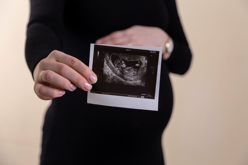
The scan will show the early pregnancy sac in the womb with the fetus within the sac. The scan is able to show this as early as 6 weeks of pregnancy.
The baby’s heartbeat is also normally visible by 6 weeks of pregnancy.
Visualising the pregnancy in the womb is reassuring. Because it confirms that it is in the right location.
How is it done?
In most cases, your doctor uses a vaginal ultrasound probe for the scan. Vaginal scanning is safe and will not harm your pregnancy in any way.
The measurements taken during the scan are the basis of an accurate EDD estimate.
Nuchal Translucency Ultrasound (Week 11–14)
Down's syndrome causes some level of learning disability. It can vary from mild to severe. Other genetic syndromes are Edwards' syndrome and Patau's syndrome.
Ultrasound scans in the first trimester assess the risk of your baby being affecting by any of these.
How is it done?
This test measures the skin fold thickness, i.e. nuchal translucency, at the back of the baby’s neck. If the neck fold is unusually thick, it may indicate that your baby may have Down's syndrome.
This test is quite accurate to detect Down Syndrome. The detection rate is 80% in the hands of experienced doctors. And the test itself is perfectly safe for your baby.
If your baby is in an optimal position, ultrasound examination after 11 weeks may be able to visualise the baby’s nasal bone. The absence of the nasal bone can be a sign of concern, which increases the risk of Down's Syndrome.
You will be informed about further testing, such as non-invasive prenatal testing, and counselling options if you receive a high risk result for Down's syndrome.
Anomaly Scans (Week 18–22)
The aim on this scan is to reassure parents-to-be that your baby will be born healthy with no congenital deformities.
This ultrasound scan screens for abnormalities in the physical structure of the baby. For example in the heart, lungs, spine, brain, long bones in legs or arms, organs in the abdomen like liver, stomach, intestines, kidneys and bladder.
Detecting of certain abnormalities will alert doctors to the possibility of genetic abnormalities in the baby. Further testing is then done to exclude these.
Most mothers and their partners enjoy the morphology ultrasound the most. This is because the view of your baby will be quite clear. You may be able to see their fingers and toes and shape of their face. This ultrasound can also find out the sex of your baby. But this depends on baby not being shy and being in a good position for the sonographer to make out if they are a boy or a girl.
How is it done?
Ultrasound screening for physical anomalies is usually done during around the fifth month or 20th week (second trimester) of your pregnancy. A sonographer or obstetrician conducts this using an advanced ultrasound machine.
The accuracy of these pregnancy ultrasound scans is about 70% in detecting all anomalies. It is impossible to provide 100% accuracy. The fetus’s structure in fact varies based on many influential factors. These include:
- maternal weight,
- reduced or increased amniotic fluid (the fluid around baby),
- abdominal trauma, and
- position of baby
The sonographer may in fact ask you to return for another ultrasound. This is because the baby is in a difficult position for them to get a clear view. With the hope that they can get a better view of your baby next time.
Third Trimester Ultrasound (32–36 weeks)
In some cases women attend ultrasounds in the third trimester.
Pregnancy Growth Ultrasound
A growth scan aims to confirm that your baby is growing well.
It is also used to determine if the amniotic fluid (water around baby) is enough. This determines the health status of your baby as well as the placenta location.
If the placenta is sitting lower in the uterus, its location may cause problems later in pregnancy or labour.
The ultrasound in this case checks to make sure that as your uterus as grown upwards in pregnancy. And that your placenta is now no longer low lying.
It is quite common for a placenta to be low lying in early pregnancy. And be in the correct position later in pregnancy.
Doppler flow studies
This ultrasound, also known as doppler scan, is a more specialised mode of ultrasound. It aims to provide more information on the blood flow status of the placenta.